October is Breast Cancer Awareness Month, a time to encourage preventative action against the most common cancer and the second leading cause of death among Canadian women.
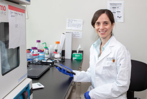
Natalia Nikolova, Electrical and Computer Engineering professor at McMaster, and Canada Research Chair in High-frequency Electromagnetics, is doing her part to combat the disease. Nikolova is working on a radar scanner for early stage breast cancer detection.
“We are developing a compact, cost-efficient and safe technology that could potentially be used in a family doctor’s office so that women can be checked as often as necessary, says Nikolova. “We have not seen this technology in clinical practice so hopefully, my team and I will be able to push this technology to revolutionize early stage breast cancer detection through mass screening programs.”
Currently, the most reliable way to find breast cancer early is screening with mammography. Though research shows this imaging technology is effective in lowering the risk of women dying from breast cancer between the ages of 50 and 69, it is expensive, not always accurate and not recommended for frequent use due to the ionization of x-ray radiation.
Radar technology employs electromagnetic waves to reveal objects that cannot be seen with the naked eye, including human tissue. The technology can be used as frequently as necessary as the radiation is nonionizing. Since compression of the scanner is minimal, the radar scanning experience would be comfortable.
“The radar scanner injects radio waves from one side of the breast, which is slightly compressed between two plates,” explains Nikolova. “The radio waves are collected on the opposite side and processed to produce images of the electrical properties of the breast tissues. This enables the differentiation of malignant from healthy tissue.”
There are a few research teams around the world pursuing similar research to Nikolova’s, but none have been approved for commercial development yet as image processing is complex and prone to errors. Nikolova’s prototype will likely be ready for clinical trial within 5 years.
“Unfortunately, this common disease has touched almost everyone in one way or another. I see my responsibility in directing my research into areas that have high social impact and improve quality of life. It is also important to inspire my students about research in science and engineering by engaging them in projects that will make a difference in people’s lives.”